We take pride in our integrity and transparency in our business dealings. Our team is friendly, can be flexible, and tries hard to accommodate our customers in all aspects of sale and support. All inquiries are answered promptly.
place an order
Fill out the information required and we will be in touch to continue the order process.
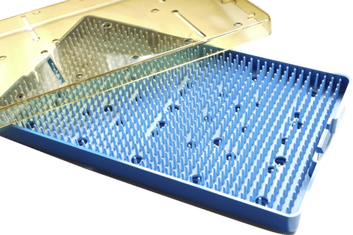
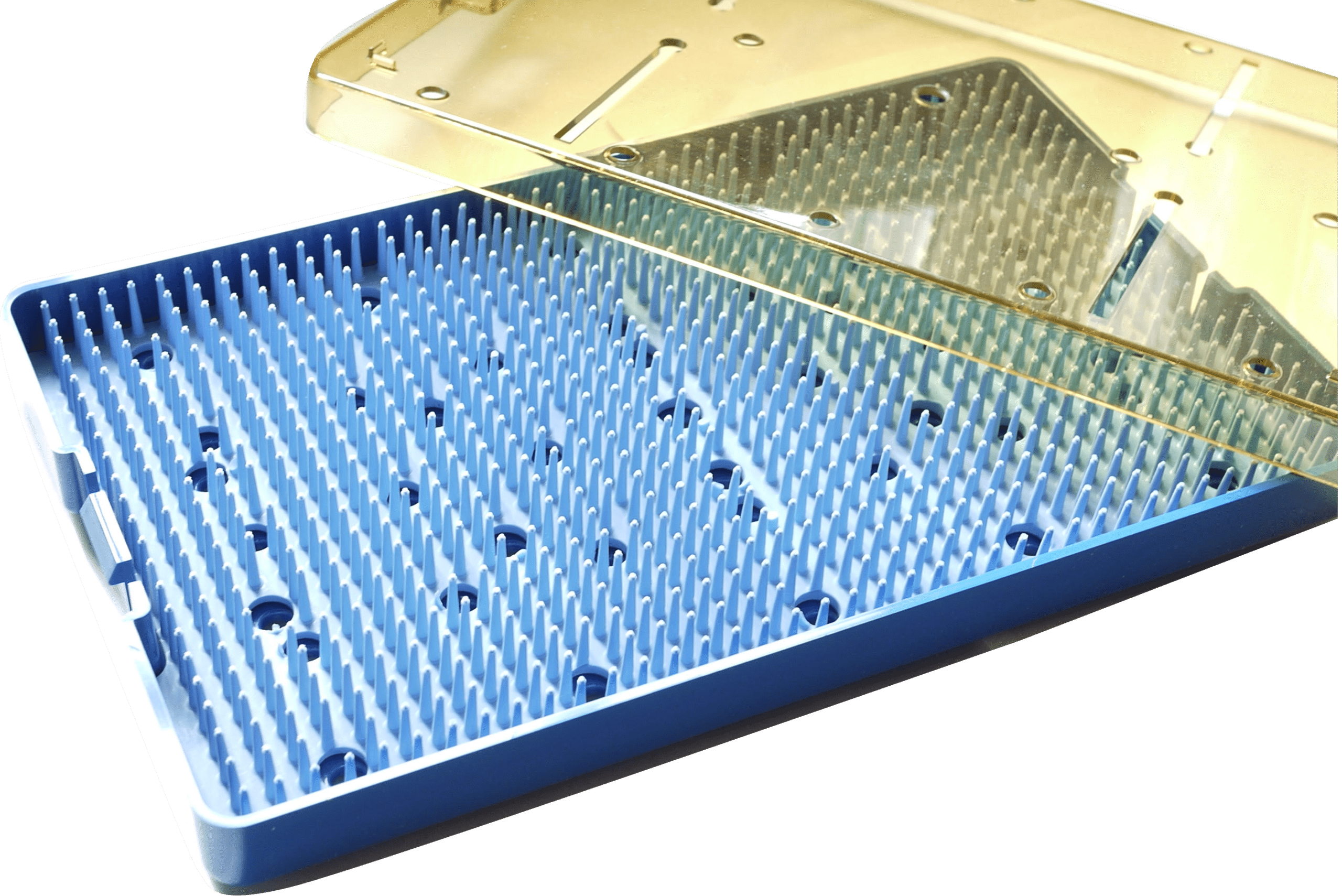
Unique, “grid” system in all bases, trays and lids, makes it easy to install the mat or insert required to protect your delicate instruments properly.
All bases have pebble-type surface to keep mats from sticking to the bases during sterilization process.
Dimensions of each tray are designed to accommodate a variety of instrument sizes and shapes.
Features Overview
High Quality Material
Durable advanced designed tray Molded from General Electric’s ULTEM® resin, using mold flow analysis to guarantee product strength, structural integrity and extended life cycle
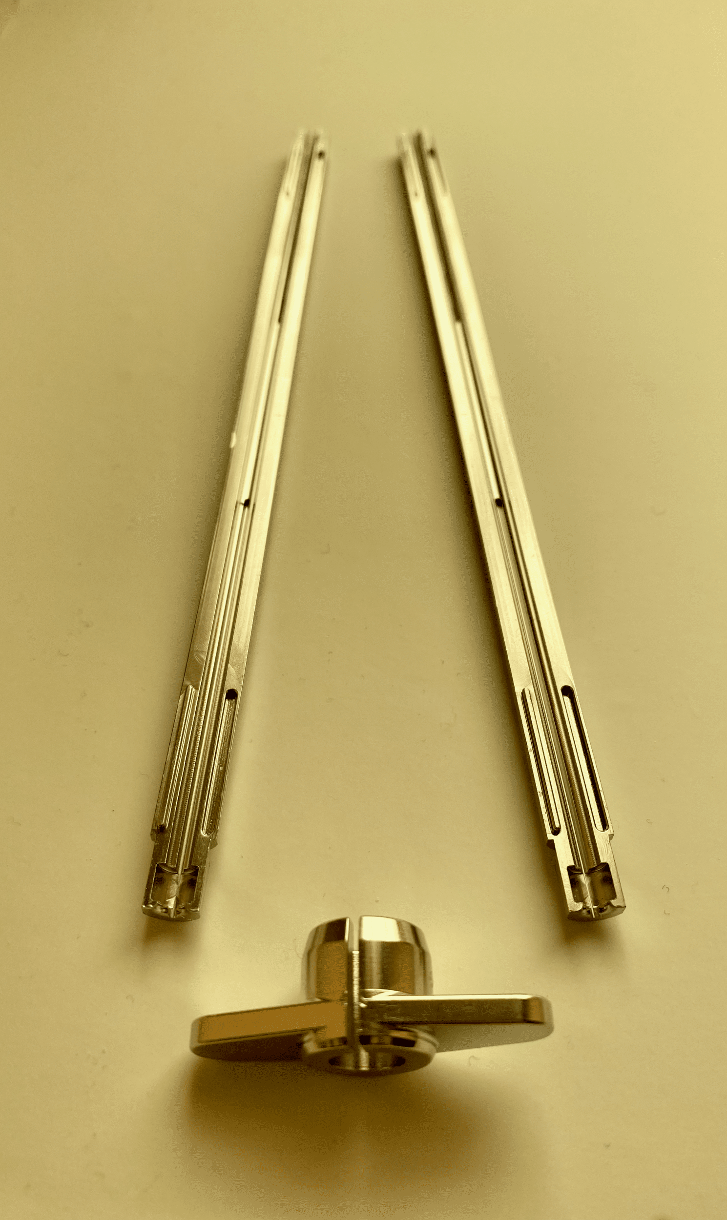
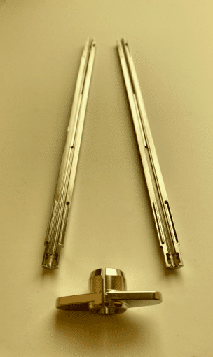
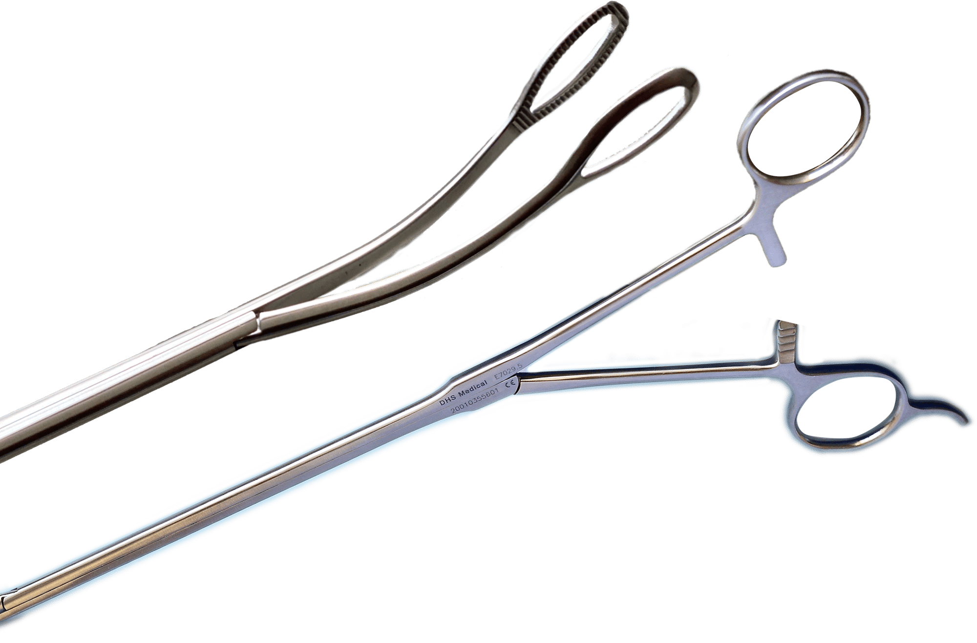
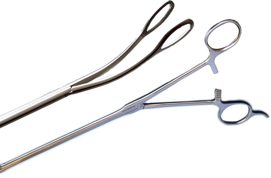
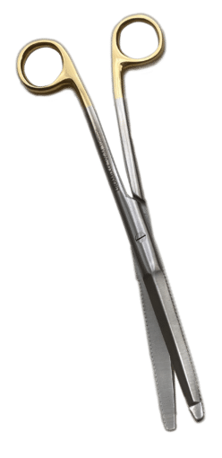
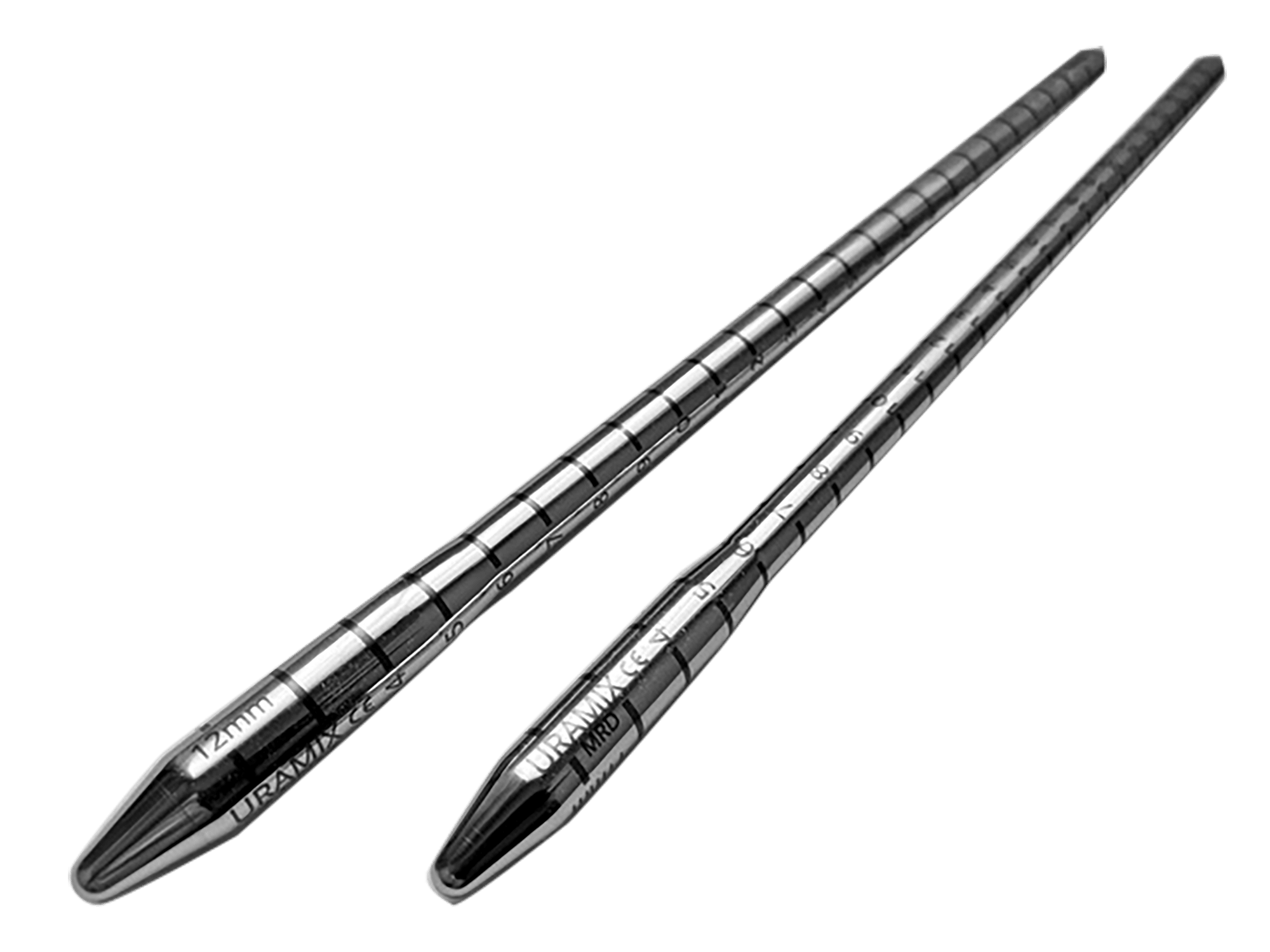
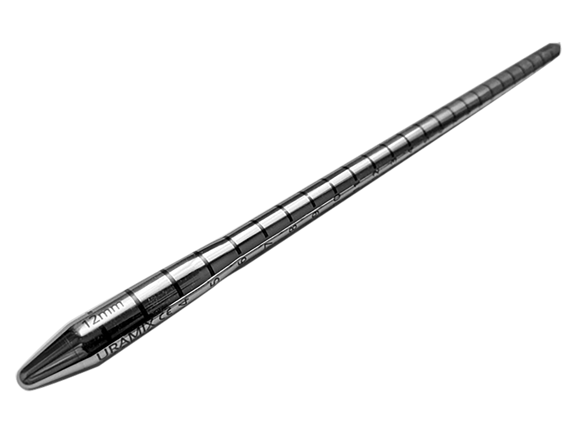
The first new needle introducer in 40 years. The Furlow Needle Introducer was made available 40 years ago. Recently, it was noted that the Furlow design, which is a closed system, can harbor proteinaceous material in its interior and thus bacteria, which can be a source of contamination at the time of the prosthesis introduction (https://doi.org/10.1038/s41443-020-0256-2). This dooms the implant and can be disastrous to the patient. The Mooreville Introducer has an open design made from two halves which come apart for easy cleaning and sterilization.
The prosthesis component introducer was improved by increasing the length of the beaks to accommodate all body habitus. They come in 3 beak lengths: 7.5 cm, 9 cm and 12 cm.
Makes introduction of prosthesis components easier with the increased beak length. It works well from both the supra-pubic approach as well as from the penoscrotal one.
Features Overview
High Quality
Made from surgical steel
Three Sizes
7.5 cm, 9 cm, and 12 cm beaks
Pricing
$299 for one
$489 for two (different sizes)
$749 for three (different sizes)
SKU # 9904
place an order
Fill out the information required and we will be in touch to continue the order process.
The NEWRC was conceived and is recommended by Dr. Steve Wilson.
Steven K. Wilson, MD, FACS, FRCS
Editor-In-Chief ISSM Video Journal of Prosthetic Urology
2007 Wilson Chair of Prosthetic Urology, U of AR
2010 St. Paul’s Medal: British Association Urologic Surgeons
2013 F Brantley Scott Award of Excellence
2017 Living Legend Award: Society Urologic Prosthetic Surgeons
Former Professor Urology: University of AR for Medical Sciences
Our Customers Said
I have used the (New Wilson Ectopic Reservoir) Clamp with great success. Very nice design.
J. Francois Eid, MD
Clinical Associate Professor of Urology Weill-Cornell Medical College Director, Advanced Urological Care, PC
Key Benefits
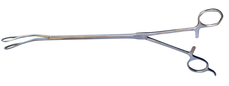
It allows placement of a prosthesis reservoir in an ectopic location with less likely bladder side effects and no visibility. The Clamp has atraumatic grasping.
The Clamp
Anatomy of a wall
Finger placement of ectopic reservoir
Reservoir in groin after finger placed ectopic reservoir
The Wilson Clamp allows the creation of a pouch in a non-visibnle position
Previous slide
Next slide
In Practice
Clamp showing how high the ectopic reservoir will be placed.
Atraumatic tip does not injure reservoir. Deaver in inguinal ring allows passage of reservoir in high abdominal wall location.
Reservoir location through a penoscrotal incision is above umbilicus.
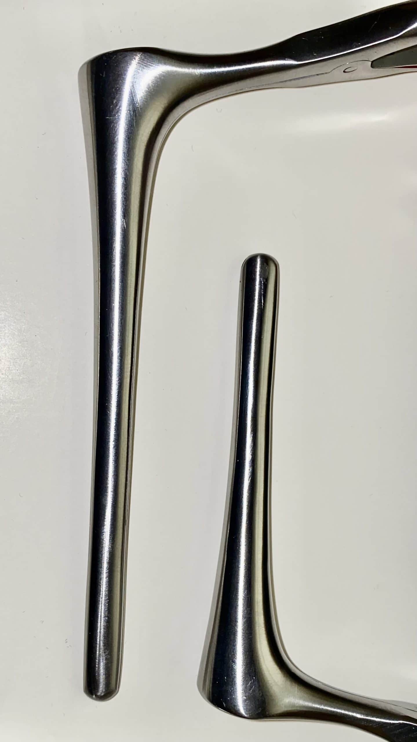
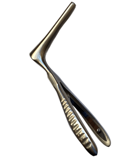
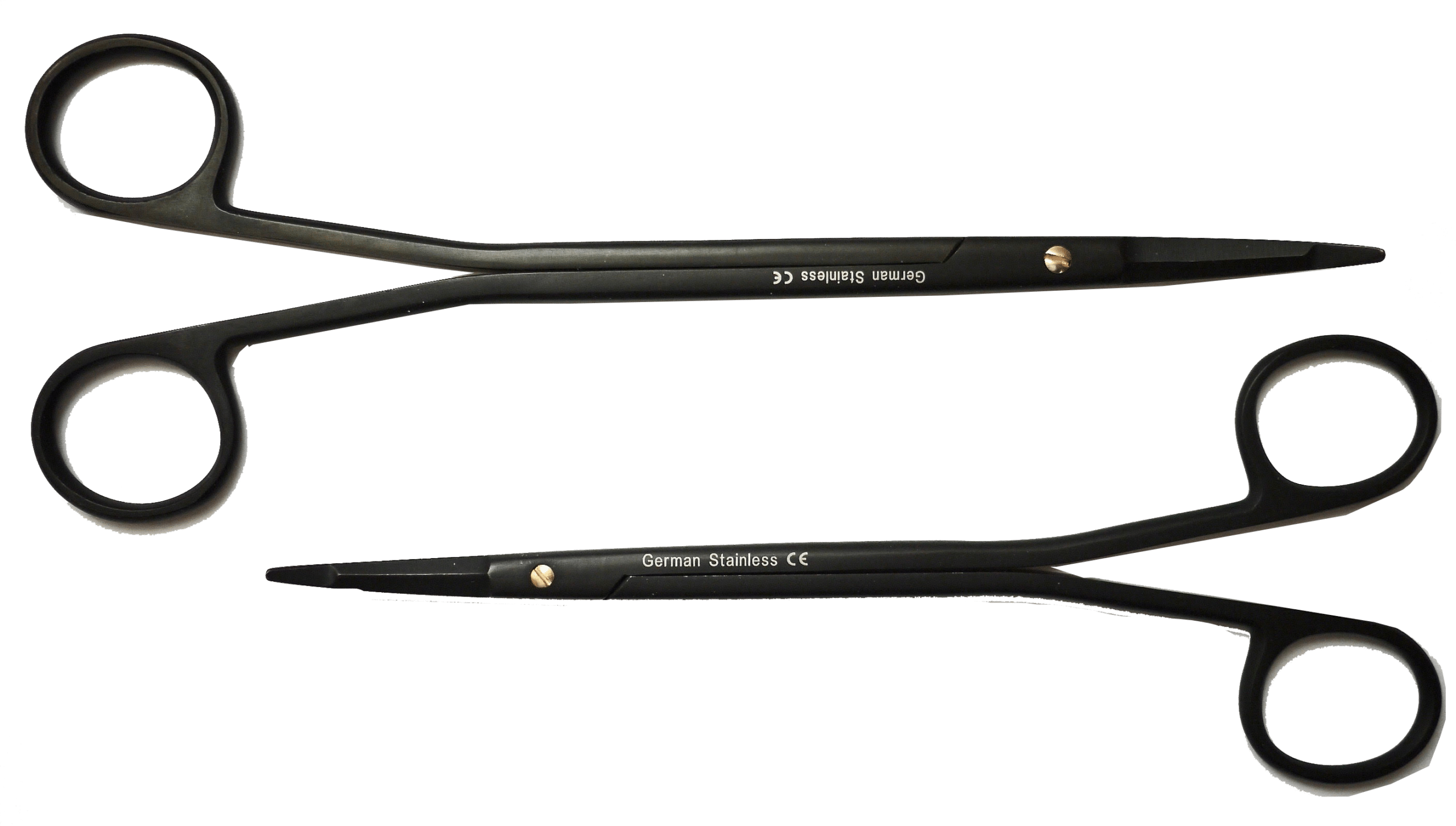
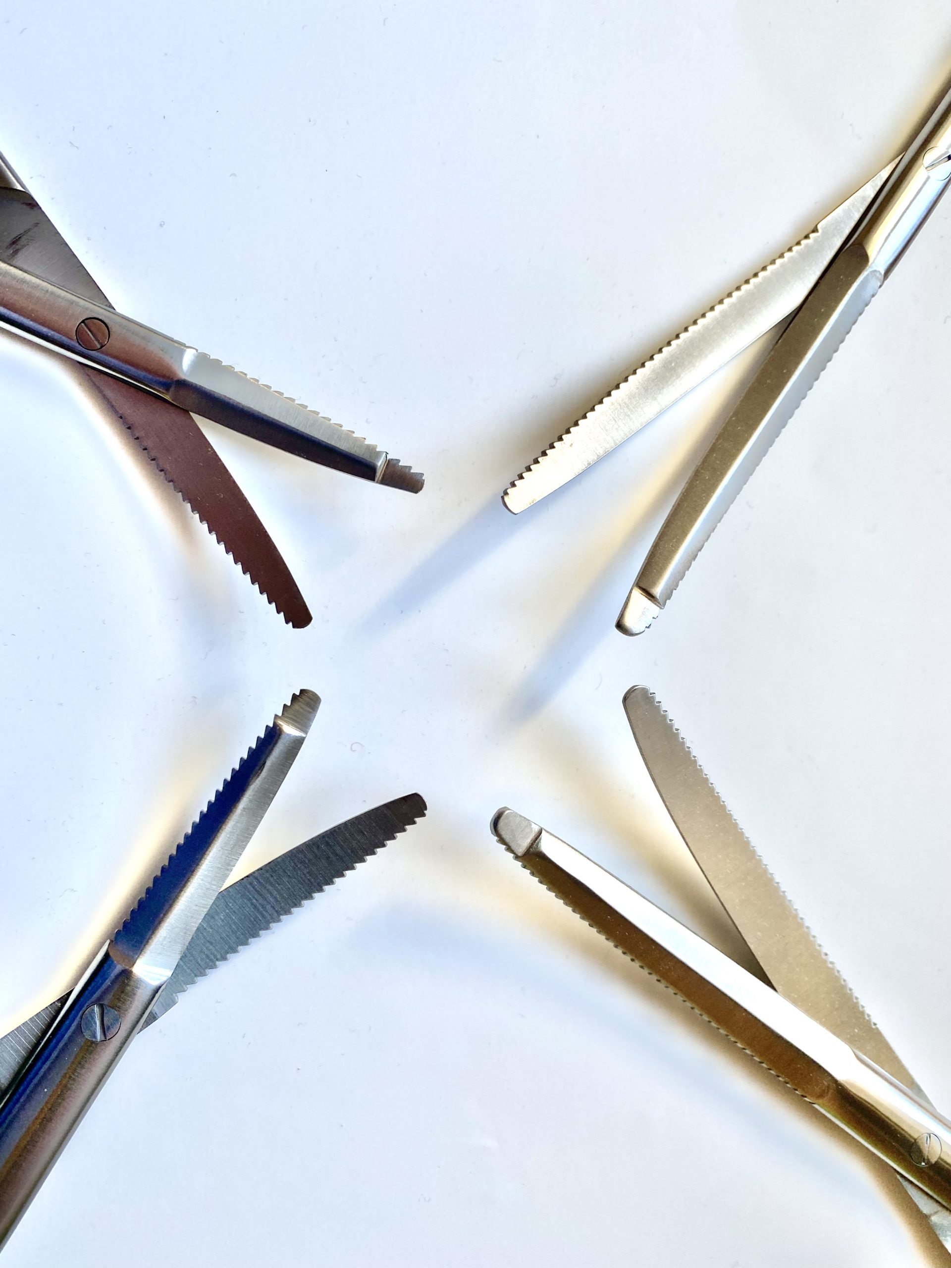
The Wilson Supercut Backward Cutting Scissor Set contains 2 scissors, a 7 and 8″ scissors for prosthetics implants in fibrotic corpora. The larger scissor is designed for the proximal corpora and the smaller one for distal work.”
The Wilson-Mooreville scissors were designed to improve the creation of space in deeply scarred corpora by the addition of lateral serrations. The scissors come in two different widths and two different tip lengths to accommodate most situations.
The TUSP was designed by Steven Ochs, a practicing urologist with Urology One in Canton, Ohio.
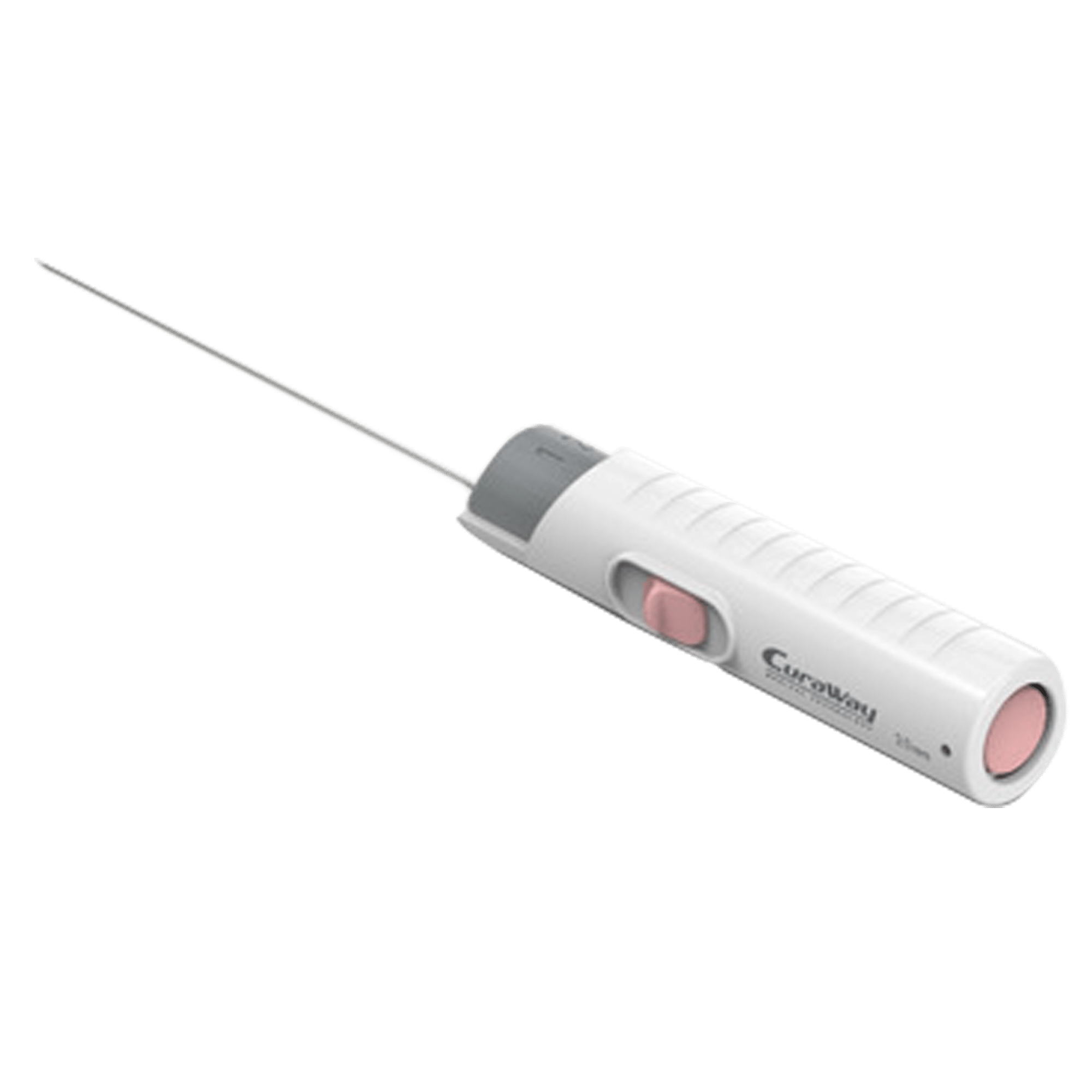
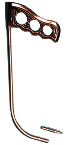
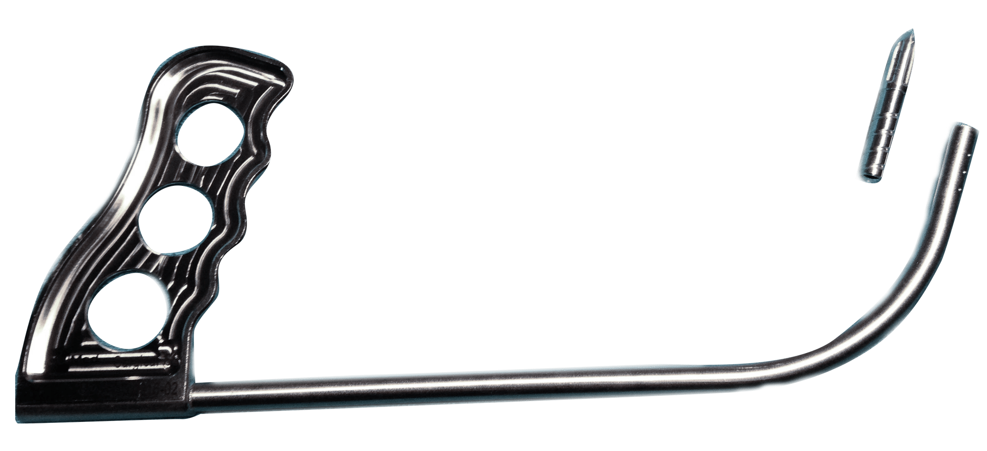
Trans-Urethral Supra-Pubic Tube Guide – The TUSP is a reusable surgical tool designed to assist the urologist in placing suprapubic tubes using the retrograde technique. It addresses the shortcoming of the Lowsley retractor to provide a safer and more efficient placement of the suprapubic tube. The TUSP features a hollow shaft that will allow the passage of up to an 18F Foley following the removal of its cannulated end piece.
Easy to use, minimally invasive, retrograde approach when antegrade approach not feasible.
Surgical Technique for ColMed TUSPTM
Step One
Use an appropriate sterile catheter. Lubricate and confirm it fits within the TUSP cannula. Lubricate and screw the trocar tip into the distal end of the cannula until flush — it is secure with minimum finger tightening.
Step Two
Place the patient in lithotomy position and prep with appropriate sterilizing solution. Perform cystoscopy to survey the anatomy of the urethra and bladder. If there is risk of urethral trauma, consider pre-placement of a guide wire (<0.038 in). Determine the optimal location for the trocar to exit the abdominal wall, and mark.
Step Three
Place the TUSP retrograde through the urethra, into the bladder. For a male, place penis on stretch. While gently angulating the handle, palpate for the tip of the device near the exit mark.
Step Four
Make an incision in the skin approximate 1 cm above the tip. While maintaining deflection on the abdominal wall with the TUSP, dissect the intervening tissue with electro-cautery until trocar emerges from the incision. Continue to push the trocar through incision about 5 cm. Remove the trocar tip and leave the cannula above the incision a few cm.
Step Five
Guide the lubricated catheter into the cannula about 25 cm (10 in) until it appears at the handle opening. If catheter buckles while advancing, push on it closer to the entry of the cannula.
Step Six
While holding the catheter at incision site, slowly withdrawal the TUSP from the urethra. Then using cystoscopic guidance retrograde through urethra, withdraw the catheter tip retrograde into the bladder by pulling externally above the incision site.
Step Six
Step Seven
Once the catheter is within the bladder lumen, inflate the balloon to recommended volume. Then gently approximate the balloon against the anterior bladder wall. Secure the catheter externally with a suture.
Previous slide
Next slide
Method of Use
Method of Use
Surgical Technique for ColMed TUSPTM
Step One
Use an appropriate sterile catheter. Lubricate and confirm it fits within the TUSP cannula. Lubricate and screw the trocar tip into the distal end of the cannula until flush — it is secure with minimum finger tightening.
Step Two
Place the patient in lithotomy position and prep with appropriate sterilizing solution. Perform cystoscopy to survey the anatomy of the urethra and bladder. If there is risk of urethral trauma, consider pre-placement of a guide wire (<0.038 in). Determine the optimal location for the trocar to exit the abdominal wall, and mark.
Step Three
Place the TUSP retrograde through the urethra, into the bladder. For a male, place penis on stretch. While gently angulating the handle, palpate for the tip of the device near the exit mark.
Step Four
Make an incision in the skin approximate 1 cm above the tip. While maintaining deflection on the abdominal wall with the TUSP, dissect the intervening tissue with electro-cautery until trocar emerges from the incision. Continue to push the trocar through incision about 5 cm. Remove the trocar tip and leave the cannula above the incision a few cm.
Step Five
Guide the lubricated catheter into the cannula about 25 cm (10 in) until it appears at the handle opening. If catheter buckles while advancing, push on it closer to the entry of the cannula.
Step Six
While holding the catheter at incision site, slowly withdrawal the TUSP from the urethra. Then using cystoscopic guidance retrograde through urethra, withdraw the catheter tip retrograde into the bladder by pulling externally above the incision site.
Step Six
Step Seven
Once the catheter is within the bladder lumen, inflate the balloon to recommended volume. Then gently approximate the balloon against the anterior bladder wall. Secure the catheter externally with a suture.
a patent-pending “soft dimple-thread," which permits the smallest size cannula (23.8 Fr) to be used with the largest sized catheter (21.5 Fr)
Pricing
$1,799 per unit
$50 for shipping
SKU # 2201
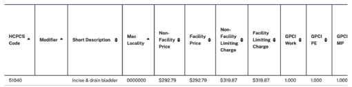
Retrograde incisional placement of SPT has higher RVU’s than punch type placement. Uramix does not endorse any particular code for billing purposes. These codes are only provided for information purposes.
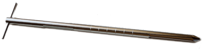
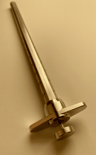
Serial dilation with Hegar or Brooks dilators has been largely abandoned by today’s prosthetic urologists…introducing the first innovation in corpora preparation in 35 years. The two preparation tools for setting up the corpora to receive a penile prosthesis have sizes: 9-11 mm and 10-12 mm. They do the work of 4 Brooks Dilators plus a measuring tool.
The Double-Bladed Cavernotome is an advancement on the original cavernotome for more challenging cases of severely scarred corpora. The cavernotome gets two cutting blades, which are sharper and rise from a beveled surface within the circumference of the dilator and the blades do not exceed this perimeter, for a precise and controlled cut or shaving action. Now available with fixed posts.
Easier and faster creation of an intracorporeal space in scarred genitalia from previous surgery or disease (i.e. – Peyronie’s, Sickle Cell). The cavernotome is great at removing bacterial film from previously infected corpora.
Play Video
Method of Use
A new double-bladed cavernotome with improved coring ability and ease of use.
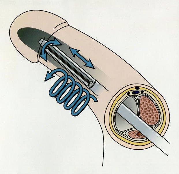
Entry to fibrotic corpora needs to be accessed by scalpel & extended both proximally & distally to allow entry to dilator
Working element should initially be directed laterally
Cutting of fibrotic tissue can be obtained longitudinally & also in a rotating shaving action
Dilators are used in succession to the desired size
Dilators can be used in nonfibrotic corpora for speedier dilation
The cavernotomes were used in nineteen patients with corporeal fibrosis. The etiology of the fibrosis was removal of previously infected prosthesis (15), extensive fibrosis from recurrent priapism secondary to sickle cell disease (2), pharmacologic injection program and subsequent Winter shunts (2). One of the patients whose prosthesis was removed for infection also had a tip erosion on the contralateral side necessitating the creation of a subcapsular space for the insertion of a new cylinder.
Surgical Technique
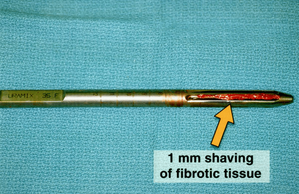
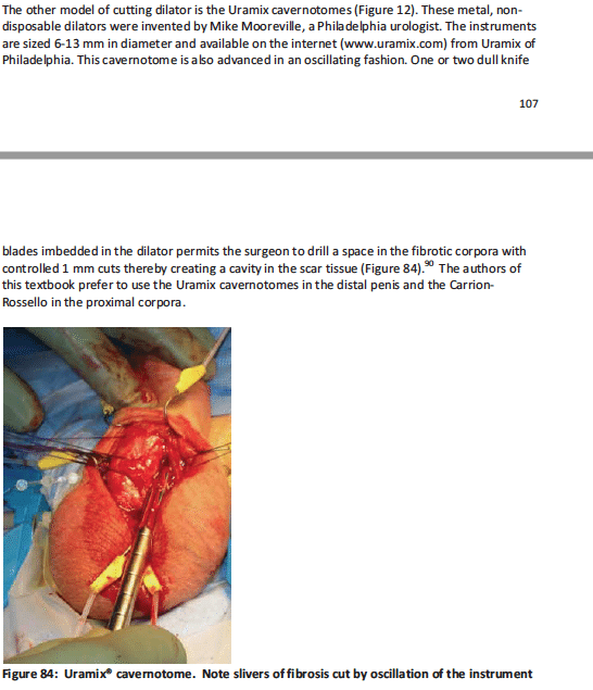
After a small corporotomy is created, the cavernotomes are introduced and moved in an oscillating motion resulting in forward advancement. If more resection is needed, rotation of the cavernotomes will create a “shaving” action which removes 1 mm strips of fibrotic tissue.
Cutting can be done both in a longitudinal, up and down, movement, or in a “drilling” rotational movement.
The largest size (13 mm) corresponds to standard cylinder diameter. Dilation to 10 mm is necessary for insertion of the Furlow tool or the Mentor NB cylinder base. Dilation to 11 mm is necessary for the insertion of the AMS CXM base.
Internal cutting of the fibrosis obviates extensive corporotomies and results in quicker procedures.
The cavernotome’s design and oscillating advancement promote safe dilation without perforation.
Cavernotome being used to dilate fibrotic corpora
All uses of the cavernotomes resulted in successful implantation of inflatable cylinders or semimalleable rods without urethral injury or corporal perforation.
Fifteen of the patients received downsized prosthesis (13 Alpha NB, 2 AMS 700 CXM).
Of the remaining four patients, three patients were implanted with Mentor Alpha 1 standard size cylinders, and 1 patient was implanted with an AMS semimalleable prosthesis.
Graft material was not required and only two patients required additional distal penile incisions for optimum cylinder tip placement.
Average operative time was 51 minutes (39-86 minutes range).
As seen in Wilson’s Perils and Pitfalls of Penile Prosthesis Surgery, Tobias S. Kohler, MD, Nikhil Gupta, MD, Steven K. Wilson, MD, 2nd edition, January 2018
Steve K. Wilson. “Reimplantation of inflatable penile prosthesis into scarred corporeal bodies”. International Journal of Impotence Research (2003) 15, Suppl 5, S125-S128.


-2")